US

Extracta DNA Prep for PCR

Quick and easy DNA extraction of PCR-ready genomic DNA
Features & Benefits
  • Simple, reagent-based system requires minimal technical skill
  • Incubation step can be carried out in 96-well PCR plates or tubes using a standard DNA thermal cycler
  • Compatible with a wide-range of clinical specimens, plant and animal tissues, and environmental samples
  • Optional stabilization buffer allows for extended storage of extracted DNA templates


Extracta DNA Prep for PCR is intended for molecular biology applications. This product is not intended for the diagnosis, prevention or treatment of a disease.

Product
Kit Size
Part Number
Price
Quantity
 
Product
Kit Size
Order Info
Price
Quantity

Extracta DNA Prep for PCR

Request Sample
Kit Size:
Part Number:
Price:
Quantity:
Kit Size:  2.5 mL
Part Number:  95091-002
Price:  $18.00
Add to Cart
Kit Size:  25 mL
Part Number:  95091-025
Price:  $164.00
Add to Cart
Kit Size:  250 mL
Part Number:  95091-250
Price:  $1,379.00
Add to Cart

Description

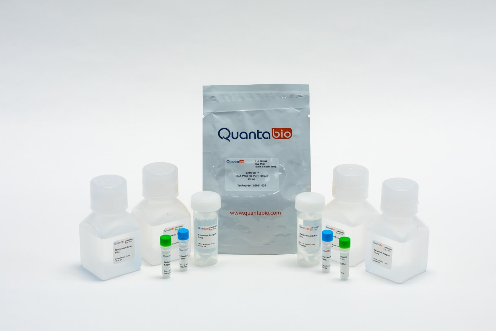
Extracta DNA Prep for PCR is a two-component reagent kit for rapid extraction of PCR-ready genomic DNA from a variety of tissues. Samples are processed in less than 30 minutes with minimal hands-on time and technical skill. Extracted genomic DNA is suitable for sensitive downstream PCR applications including end-point PCR, High Resolution Melt Analysis (HRM) and quantitative real-time PCR (qPCR) without requiring any additional clean-up. In addition, the extracted DNA may be
used in multiplexed PCR applications such as transgene or knock-out analyses. Tissue extractions can be done in tubes, plates or deep-well blocks to allow for adaptation to workflow and automation on liquid-handling workstations.

Details

Details

Contents
  • Extraction Reagent (1 x 25 mL or 2 x 125 mL)
  • Stabilization Buffer (1 x 25 mL or 2 x 125 mL)
Customer Testimonials

Customer Testimonials

Extracta DNA Prep for PCR
"gDNA can be extracted from as low as one single cell. I used this kit on marine ciliates and was able to get few ng of DNA for PCR amplification."
Fellow | Texas A&M University
Extracta DNA Prep for PCR
"This kit is faster than others that I used before. I can perform the PCR with lower amount of input, perfect for small ear punches instead of cut the tail. Also, the PCR results are more clean."
Scientist | CABIMER
Extracta DNA Prep for PCR
"It worked very well for our needs. It is easy to use and gave strong, clean bands in PCR when used for genotyping."
Fellow | WEILL Cornell Medical College
Extracta DNA Prep for PCR

"I needed to extract DNA from fungal tissue for one of our teaching labs. It worked amazing for further PCR reactions."


Andres P. | University Lab Coordinator
Extracta DNA Prep for PCR - Tissue

"Extracta DNA Prep for PCR  is one of the best, fastest and easiest to use reagent for genomic DNA isolation from human cells."

Scientist | Deutsches Krebsforschungszentrum (DKFZ)
Related Products

Details

Contents
  • Extraction Reagent (1 x 25 mL or 2 x 125 mL)
  • Stabilization Buffer (1 x 25 mL or 2 x 125 mL)

Performance Data

Resources

Customer Profile Stories

Flyers

Product Manuals

Application Notes

CofA (PSFs)

Click here to see all CofA (PSFs)

SDSs

Publications

A rapid, cost-effective, colorimetric LAMP assay (CLASS) for detecting invasive malaria vector, Anopheles stephensi
Cristina Rafferty - 2024
Abstract
Anopheles stephensi, an invasive malaria vector in Africa, has the potential to impact the landscape of malaria on the continent, threatening to put an additional 126 million people per year at risk of malaria, largely in peri-urban/urban areas. To accelerate the early detection and rapid response to An. stephensi and ensure no gains made in malaria control and elimination are lost, it is critical to confirm the presence of the species and the geographic extent of its spread to inform control. However, morphological identification may be misinterpreted if specimens are damaged and existing molecular species confirmation assays require specialized laboratory equipment and training and may be challenging to interpret, requiring additional sequencing confirmation. A colorimetric rapid loop-mediated isothermal amplification (LAMP) assay for molecular An. stephensi species identification was developed and optimized. The colorimetric assay requires only a heat source and reagents and can be used with or without DNA extraction resulting in positive color change in 30-35 minutes. To determine analytical sensitivity, a 1:10 dilution series of the DNA extract was conducted showing 100% assay sensitivity down to 0.003 nanograms. To determine specificity, three different An. stephensi laboratory strains (STE2, SDA 500, UCI), 8 other Anopheles mosquito species, and Aedes aegypti were compared, and the results indicated 100% specificity across these species. To determine use without the need for DNA extraction, samples evaluated included a single mosquito leg, whole adult or larval mosquitoes, and pooled DNA extract from several mosquito species. A total of 1687 individual reactions were tested during optimization and all LAMP assay results were compared against the conventional PCR assay and confirmed through Sanger sequencing. To validate the optimized assay on wild caught specimens, DNA extracted from 12 wild caught, sequence-confirmed An. stephensi from Marsabit, Kenya, were tested and the colorimetric assay was accurate in identifying all of the specimens as An. stephensi. The assay described presents an opportunity to accelerate An. stephensi molecular identification in new and existing locations in Africa, within its endemic range, and globally. These findings present a simple, rapid, unique alternative to existing PCR and sequencing-based An. stephensi species identification and confirmation strategies. With additional field validation studies, molecular screening tools like the colorimetric LAMP-based An. stephensi species identification (CLASS) assay fill an important gap of rapid confirmation of this invasive vector and presents an ideal opportunity to better understand the spread of the species in Africa and other recently invaded areas, thus accelerating a response to mitigate its long-term impacts on malaria on the continent.
Evaluating the impact of GPER1 and CCR5 during ZIKV infection in pregnant mice
J. Walker Lee, Jr. - 2023
Abstract
Zika virus (ZIKV) is the most recent flavivirus to cause a large-scale outbreak worldwide. Unlike other flaviviruses, ZIKV can be vertically-transmitted from mother to fetus. Infection of pregnant individuals with ZIKV, irrespective of symptom development, can lead to microcephaly and other congenital malformations, as well as fetal loss, stillbirth, and preterm birth. In this thesis, we evaluate the role of two genes: GPER1 (Chapter 3), an estrogen receptor recently identified to function in protecting fetuses against type I Interferon-mediated damage, and CCR5 (Chapter 4), a chemokine receptor known to control flavivirus damage to the CNS. To do this, we have backcrossed GPER1 and CCR5 knockout mice to the humanized STAT2 knock in mice (hSTAT2KI), a mouse strain recently generated that is more permissive to ZIKV infection. Timed pregnancies were conducted, and mice were infected at E6.5 to evaluate ZIKV pathogenesis in the presence or absence of GPER1 or CCR5. Pregnancies were stopped at E13.5 (7 days post infection) or allowed to proceed to term to evaluate litter size, pup survival, as well as a wide range of physical, sensory, and motor developmental milestone abnormalities. For GPER1, we observed a significant protective effect in regard to litter size and survival following in utero ZIKV exposure, but no effect with regards to any developmental outcomes. For CCR5, we observed significant protective effects on congenital survival and several aspects of development, with the absence of CCR5 correlating with a host of physical and motor developmental delays following in utero ZIKV exposure. Overall, our studies suggest that both GPER1 and CCR5 play roles in the protection of fetal health in response to ZIKV infection, with GPER1 functioning primarily to protect prior to birth, and CCR5 functioning more significantly post birth.
ER and SOCE Ca2+ signals are not required for directed cell migration in human microglia
Alberto Granzotto - 2024
Abstract
The central nervous system (CNS) is constantly surveilled by microglia, highly motile and dynamic cells deputed to act as the first line of immune defense in the brain and spinal cord. Alterations in the homeostasis of the CNS are detected by microglia that respond by migrating toward the affected area. Understanding the mechanisms controlling directed cell migration of microglia is crucial to dissect their responses to neuroinflammation and injury. We used a combination of pharmacological and genetic approaches to explore the involvement of calcium (Ca2+) signaling in the directed migration of induced pluripotent stem cell (iPSC)-derived microglia challenged with a purinergic stimulus. This approach mimics cues originating from injury of the CNS. Unexpectedly, simultaneous imaging of microglia migration and intracellular Ca2+ changes revealed that this phenomenon does not require Ca2+ signals generated from the endoplasmic reticulum (ER) and store-operated Ca2+ entry (SOCE) pathways. Instead, we find evidence that human microglial chemotaxis to purinergic signals is mediated by cyclic AMP in a Ca2+ -independent manner. These results challenge prevailing notions, with important implications in neurological conditions characterized by perturbation in Ca2+ homeostasis.
Smooth muscle–derived adventitial progenitor cells direct atherosclerotic plaque composition complexity in a Klf4-dependent manner
Allison M. Dubner - 2023
Abstract
We previously established that vascular smooth muscle–derived adventitial progenitor cells (AdvSca1-SM) preferentially differentiate into myofibroblasts and contribute to fibrosis in response to acute vascular injury. However, the role of these progenitor cells in chronic atherosclerosis has not been defined. Using an AdvSca1-SM cell lineage tracing model, scRNA-Seq, flow cytometry, and histological approaches, we confirmed that AdvSca1-SM–derived cells localized throughout the vessel wall and atherosclerotic plaques, where they primarily differentiated into fibroblasts, smooth muscle cells (SMC), or remained in a stem-like state. Krüppel-like factor 4 (Klf4) knockout specifically in AdvSca1-SM cells induced transition to a more collagen-enriched fibroblast phenotype compared with WT mice. Additionally, Klf4 deletion drastically modified the phenotypes of non–AdvSca1-SM–derived cells, resulting in more contractile SMC and atheroprotective macrophages. Functionally, overall plaque burden was not altered with Klf4 deletion, but multiple indices of plaque composition complexity, including necrotic core area, macrophage accumulation, and fibrous cap thickness, were reduced. Collectively, these data support that modulation of AdvSca1-SM cells through KLF4 depletion confers increased protection from the development of potentially unstable atherosclerotic plaques.
The Use of Saliva Samples to Test for Congenital Cytomegalovirus Infection in Newborns: Examination of False-Positive Samples Associated with Donor Milk Use
Whitney Wunderlich - 2023
Abstract
A universal screening research study was conducted in six hospitals to identify the clinical sensitivity of polymerase chain reaction (PCR) testing on newborn dried blood spots (DBSs) versus saliva specimens for the diagnosis of congenital cytomegalovirus (cCMV). CMV DNA positive results from DBSs or saliva were confirmed with urine testing. Findings of several false-positive (FP) saliva PCR results prompted an examination of a possible association with donor milk. Documentation of the frequency of positive saliva results, including both true-positive (TP) and FP status from clinical confirmation, occurred. The frequency of donor milk use was compared for TP and FP cases. Of 22,079 participants tested between 2016 and 2022, 96 had positive saliva results, 15 were determined to be FP, 79 TP, and 2 were excluded for incomplete clinical evaluation. Newborn donor milk use was identified for 18 (19.14%) of all the positive saliva screens. Among the 15 FPs, 11 (73.33%) consumed donor milk compared to 7 of the 79 TPs (8.8%) (OR 28.29, 95% CI 7.10–112.73, p < 0.001). While milk bank Holder pasteurization inactivates CMV infectivity, CMV DNA may still be detectable. Due to this possible association, screening programs that undertake testing saliva for CMV DNA may benefit from documenting donor milk use as a potential increased risk for FP results.
Amphiregulin couples IL1RL1+ regulatory T cells and cancer-associated fibroblasts to impede antitumor immunity
Runzi Sun - 2023
Abstract
Regulatory T (Treg) cells and cancer-associated fibroblasts (CAFs) jointly promote tumor immune tolerance and tumorigenesis. The molecular apparatus that drives Treg cell and CAF coordination in the tumor microenvironment (TME) remains elusive. Interleukin 33 (IL-33) has been shown to enhance fibrosis and IL1RL1+ Treg cell accumulation during tumorigenesis and tissue repair. We demonstrated that IL1RL1 signaling in Treg cells greatly dampened the antitumor activity of both IL-33 and PD-1 blockade. Whole tumor single-cell RNA sequencing (scRNA-seq) analysis and blockade experiments revealed that the amphiregulin (AREG)–epidermal growth factor receptor (EGFR) axis mediated cross-talk between IL1RL1+ Treg cells and CAFs. We further demonstrated that the AREG/EGFR axis enables Treg cells to promote a profibrotic and immunosuppressive functional state of CAFs. Moreover, AREG mAbs and IL-33 concertedly inhibited tumor growth. Our study reveals a previously unidentified AREG/EGFR-mediated Treg/CAF coupling that controls the bifurcation of fibroblast functional states and is a critical barrier for cancer immunotherapy.
Plaque-associated human microglia accumulate lipid droplets in a chimeric model of Alzheimer’s disease
Christel Claes - 2021
Abstract
Background Disease-associated microglia (DAMs), that surround beta-amyloid plaques, represent a transcriptionally-distinct microglial profile in Alzheimer’s disease (AD). Activation of DAMs is dependent on triggering receptor expressed on myeloid cells 2 (TREM2) in mouse models and the AD TREM2-R47H risk variant reduces microglial activation and plaque association in human carriers. Interestingly, TREM2 has also been identified as a microglial lipid-sensor, and recent data indicates lipid droplet accumulation in aged microglia, that is in turn associated with a dysfunctional proinflammatory phenotype. However, whether lipid droplets (LDs) are present in human microglia in AD and how the R47H mutation affects this remains unknown. Methods To determine the impact of the TREM2 R47H mutation on human microglial function in vivo, we transplanted wild-type and isogenic TREM2-R47H iPSC-derived microglial progenitors into our recently developed chimeric Alzheimer mouse model. At 7 months of age scRNA-seq and histological analyses were performed. Results Here we report that the transcriptome of human wild-type TREM2 and isogenic TREM2-R47H DAM xenografted microglia (xMGs), isolated from chimeric AD mice, closely resembles that of human atherosclerotic foam cells. In addition, much like foam cells, plaque-bound xMGs are highly enriched in lipid droplets. Somewhat surprisingly and in contrast to a recent in vitro study, TREM2-R47H mutant xMGs exhibit an overall reduction in the accumulation of lipid droplets in vivo. Notably, TREM2-R47H xMGs also show overall reduced reactivity to plaques, including diminished plaque-proximity, reduced CD9 expression, and lower secretion of plaque-associated APOE. Conclusions Altogether, these results indicate lipid droplet accumulation occurs in human DAM xMGs in AD, but is reduced in TREM2-R47H DAM xMGs, as it occurs secondary to TREM2-mediated changes in plaque proximity and reactivity.
The entomological impact of passive metofluthrin emanators against indoor Aedes aegypti: A randomized field trial
Gregor J. Devine - 2021
Abstract
Background In the absence of vaccines or drugs, insecticides are the mainstay of Aedes-borne disease control. Their utility is challenged by the slow deployment of resources, poor community compliance and inadequate household coverage. Novel application methods are required. Methodology and principal findings A 10% w/w metofluthrin “emanator” that passively disseminates insecticide from an impregnated net was evaluated in a randomized trial of 200 houses in Mexico. The devices were introduced at a rate of 1 per room and replaced at 3-week intervals. During each of 7 consecutive deployment cycles, indoor resting mosquitoes were sampled using aspirator collections. Assessments of mosquito landing behaviours were made in a subset of houses. Pre-treatment, there were no differences in Aedes aegypti indices between houses recruited to the control and treatment arms. Immediately after metofluthrin deployment, the entomological indices between the trial arms diverged. Averaged across the trial, there were significant reductions in Abundance Rate Ratios for total Ae. aegypti, female abundance and females that contained blood meals (2.5, 2.4 and 2.3-times fewer mosquitoes respectively; P<0.001). Average efficacy was 60.2% for total adults, 58.3% for females, and 57.2% for blood-fed females. The emanators also reduced mosquito landings by 90% from 12.5 to 1.2 per 10-minute sampling period (P<0.05). Homozygous forms of the pyrethroid resistant kdr alleles V410L, V1016L and F1534C were common in the target mosquito population; found in 39%, 24% and 95% of mosquitoes collected during the trial. Conclusions/Significance This is the first randomized control trial to evaluate the entomological impact of any volatile pyrethroid on urban Ae. aegypti. It demonstrates that volatile pyrethroids can have a sustained impact on Ae. aegypti population densities and human-vector contact indoors. These effects occur despite the presence of pyrethroid-resistant alleles in the target population. Formulations like these may have considerable utility for public health vector control responses.
The relationship between insecticide resistance, mosquito age and malaria prevalence in Anopheles gambiae s.l. from Guinea
Emma Collins - 2019
Abstract
Insecticide resistance across sub-Saharan Africa may impact the continued effectiveness of malaria vector control. We investigated the association between carbamate and pyrethroid resistance with Anopheles gambiae s.l. parity, Plasmodium falciparum infection, and molecular insecticide resistance mechanisms in Guinea. Pyrethroid resistance was intense, with field populations surviving ten times the insecticidal concentration required to kill susceptible individuals. The L1014F kdr-N1575Y haplotype and I1527T mutation were significantly associated with mosquito survival following permethrin exposure (Prevalence Ratio; PR = 1.92, CI = 1.09–3.37 and PR = 2.80, CI = 1.03–7.64, respectively). Partial restoration of pyrethroid susceptibility following synergist pre-exposure suggests a role for mixed-function oxidases. Carbamate resistance was lower and significantly associated with the G119S Ace-1 mutation. Oocyst rates were 6.8% and 4.2% among resistant and susceptible mosquitoes, respectively; survivors of bendiocarb exposure were significantly more likely to be infected. Pyrethroid resistant mosquitoes had significantly lower parity rates than their susceptible counterparts (PR = 1.15, CI = 1.10–1.21). Our findings emphasize the need for additional studies directly assessing the influence of insecticide resistance on mosquito fitness.
Click here to see all Publications

Customer Testimonials

Extracta DNA Prep for PCR
"gDNA can be extracted from as low as one single cell. I used this kit on marine ciliates and was able to get few ng of DNA for PCR amplification."
Fellow | Texas A&M University
Extracta DNA Prep for PCR
"This kit is faster than others that I used before. I can perform the PCR with lower amount of input, perfect for small ear punches instead of cut the tail. Also, the PCR results are more clean."
Scientist | CABIMER
Extracta DNA Prep for PCR
"It worked very well for our needs. It is easy to use and gave strong, clean bands in PCR when used for genotyping."
Fellow | WEILL Cornell Medical College
Extracta DNA Prep for PCR

"I needed to extract DNA from fungal tissue for one of our teaching labs. It worked amazing for further PCR reactions."


Andres P. | University Lab Coordinator
Extracta DNA Prep for PCR - Tissue

"Extracta DNA Prep for PCR  is one of the best, fastest and easiest to use reagent for genomic DNA isolation from human cells."

Scientist | Deutsches Krebsforschungszentrum (DKFZ)

Welcome to the Quantabio webshop!

Please complete the new user account registration form

Once your account is setup, you'll be able to purchase any of the products on our site at any time with next day delivery for in stock products.

Product Finder

Select Your Assay

Starting Template

Assay Format

Detection Chemistry

Multiplexing (more than 3 targets)

Is gene-specific priming (GSP) required?

What current Reverse Transcriptase or cDNA kit are you using?

Select the group which contains your real-time PCR cycler

  • Applied Biosystems 7500
  • Applied Biosystems 7500 Fast
  • Stratagene Mx3000P®
  • Stratagene Mx3005P™
  • Stratagene Mx4000™
  • Applied Biosystems ViiA 7
  • Applied Biosystems QuantStudio™
  • Agilent AriaMx
  • Douglas Scientific IntelliQube®
  • Applied Biosystems 5700
  • Applied Biosystems 7000
  • Applied Biosystems 7300
  • Applied Biosystems 7700
  • Applied Biosystems 7900
  • Applied Biosystems 7900HT
  • Applied Biosystems 7900 HT Fast
  • Applied Biosystems StepOne™
  • Applied Biosystems StepOnePlus™
  • Quantabio Q
  • BioRad CFX
  • Roche LightCycler 480
  • QIAGEN Rotor-Gene Q
  • Azure Cielo 6
  • Azure Cielo 3
  • Other
  • BioRad iCycler iQ™
  • BioRad MyiQ™
  • BioRad iQ™5

Choose your application from the categories below

Products

I give Quantabio or an authorized Quantabio distributor permission to contact me for product updates and news.
* Required information